Questa pagina non è una pagina ufficiale dell'app o del suo sviluppatore, ma una pubblicazione editoriale indipendente creata a scopo informativo e di commento. Salvo espressa indicazione contraria, né l'app né il suo sviluppatore sono affiliati, approvati, sponsorizzati, autorizzati o altrimenti ufficialmente collegati a MWM, Apple, Google Play, all'editore dell'app o allo sviluppatore dell'app, e nulla in questa pagina implica che l'app sia stata sviluppata utilizzando i servizi di MWM. Tutti i marchi, loghi, screenshot e altri contenuti rimangono di proprietà dei rispettivi proprietari.

teamLab Body Pro 3d anatomy
Migliora la tua pratica medica con il primo modello 3D al mondo basato su oltre 10 anni di dati MRI. Visualizza il movimento articolare dinamico, esegui sezioni trasversali personalizzate ed esplora la struttura umana con precisione di livello chirurgico.
Download
108K+Valutazione
Valutazioni totali
600Editore
Categoria
MedicalLingue
1Ultima versione
1.3.5Dimensione
300.9 MBData di lancio
6 nov 2022Lo Standard d'Oro nell'Anatomia 3D
Sostituisci i libri di testo statici con il modello 3D più accurato al mondo, derivato da 10 anni di ricerca MRI dal vivo per supportare la precisione clinica e l'apprendimento medico avanzato.
Biomeccanica delle Articolazioni Viventi
Padroneggia la kinesiologia con le prime animazioni 3D al mondo del movimento articolare dal vivo, fornendo una rappresentazione più accurata rispetto ai tradizionali studi su cadavere.
Sezionamento Clinico Trasversale
Genera istantaneamente piani sagittali, frontali o arbitrari personalizzati, progettati per assistere nella diagnosi ecografica e nella complessa pianificazione chirurgica.
Gli screenshot e la descrizione seguenti provengono direttamente dall'elenco ufficiale dello store dell'app e sono di proprietà dello sviluppatore.
App Store
Screenshot
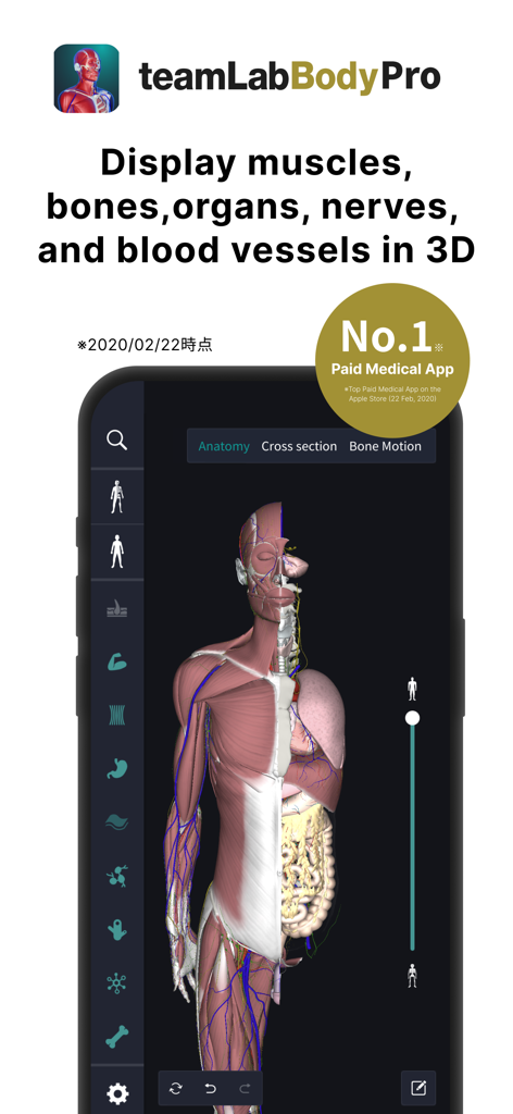
Modello anatomico umano 3D che mostra muscoli e organi nell'app teamLab Body Pro
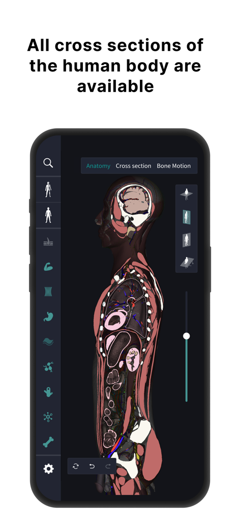
Vista di sezione trasversale del corpo umano nell'app di anatomia 3D teamLab Body Pro.
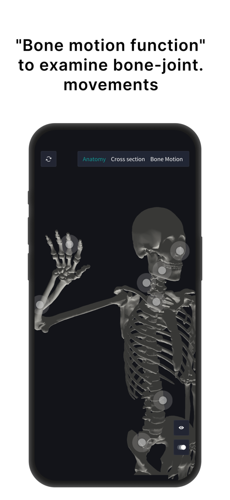
Modello di scheletro umano 3D in teamLab Body Pro che mostra punti interattivi di movimento articolare
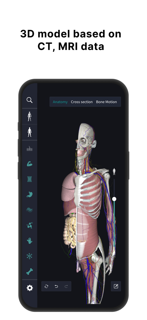
Modello anatomico umano 3D che mostra organi e muscoli basati su dati MRI
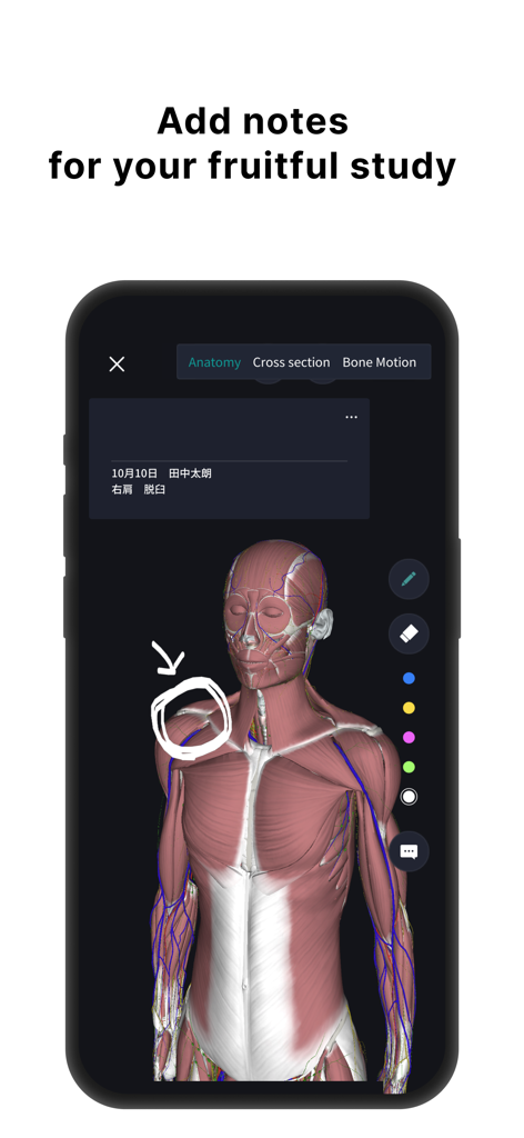
Modello anatomico umano 3D con strati muscolari e annotazioni disegnate a mano per lo studio medico.
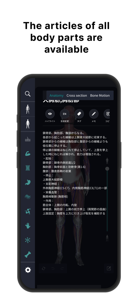
Descrizioni anatomiche dettagliate e interfaccia del modello umano 3D.

Recensioni degli utenti per l'app di anatomia 3D teamLab Body Pro che evidenziano i suoi benefici per professionisti sanitari e studenti
Descrizione
Download
App Simili
App con funzionalità ed esperienza simili

Acupuncture Points
Miridia Technology, Inc.

Anatomy by M&M
Muscle & Motion LTD

みんなde心電図
Ryoma Kamei

Histo!
John Ash

Posture by M&M
Muscle & Motion LTD

NYSORA VetRA
NYSORA Inc

Physiotutors
Physiotutors

BLOCKJOCKS
BLOCKJOCKS.COM LLC

Eastland Herb–Chinese Medicine
Eastland Press Inc.
Questa pagina non è una pagina ufficiale dell'app o del suo sviluppatore, ma una pubblicazione editoriale indipendente creata a scopo informativo e di commento. Salvo espressa indicazione contraria, né l'app né il suo sviluppatore sono affiliati, approvati, sponsorizzati, autorizzati o altrimenti ufficialmente collegati a MWM, Apple, Google Play, all'editore dell'app o allo sviluppatore dell'app, e nulla in questa pagina implica che l'app sia stata sviluppata utilizzando i servizi di MWM. Tutti i marchi, loghi, screenshot e altri contenuti rimangono di proprietà dei rispettivi proprietari.