Esta página não é uma página oficial do aplicativo ou de seu desenvolvedor, mas uma publicação editorial independente criada para fins informativos e de comentário. Salvo indicação expressa em contrário, nem o aplicativo nem seu desenvolvedor são afiliados, endossados, patrocinados, autorizados ou oficialmente conectados à MWM, Apple, Google Play, ao editor do aplicativo ou ao desenvolvedor do aplicativo, e nada nesta página implica que o aplicativo foi desenvolvido utilizando os serviços da MWM. Todas as marcas comerciais, logotipos, capturas de tela e outros conteúdos permanecem propriedade de seus respectivos proprietários.

teamLab Body Pro 3d anatomy
Eleve sua prática médica com o primeiro modelo 3D do mundo baseado em mais de 10 anos de dados de ressonância magnética. Visualize movimentos dinâmicos das articulações, realize secções transversais personalizadas e explore a estrutura humana com precisão de nível cirúrgico.
Downloads
108K+Avaliação
Total de avaliações
600Editor
Categoria
MedicalIdiomas
1Última versão
1.3.5Tamanho
300.9 MBData de lançamento
6 de nov. de 2022O Padrão Ouro em Anatomia 3D
Substitua livros estáticos pelo modelo 3D mais preciso do mundo, derivado de 10 anos de pesquisa de ressonância magnética ao vivo para apoiar a precisão clínica e o aprendizado médico avançado.
Biomecânica Articular Viva
Domine a cinesiologia com as primeiras animações 3D de movimento articular ao vivo do mundo, fornecendo uma representação mais precisa do que estudos cadavéricos tradicionais.
Secções Transversais Clínicas
Gere instantaneamente planos sagitais, frontais ou arbitrários personalizados, projetados para auxiliar no diagnóstico por ultrassom e no planejamento cirúrgico complexo.
As capturas de tela e a descrição a seguir são provenientes diretamente da listagem oficial da loja do aplicativo e são propriedade do desenvolvedor.
App Store
Capturas
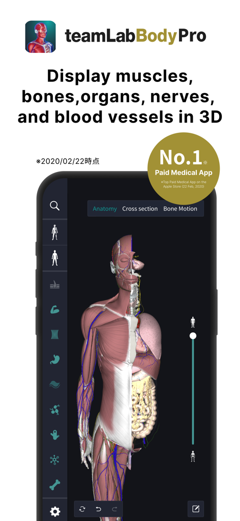
Modelo 3D de anatomia humana mostrando músculos e órgãos no aplicativo teamLab Body Pro
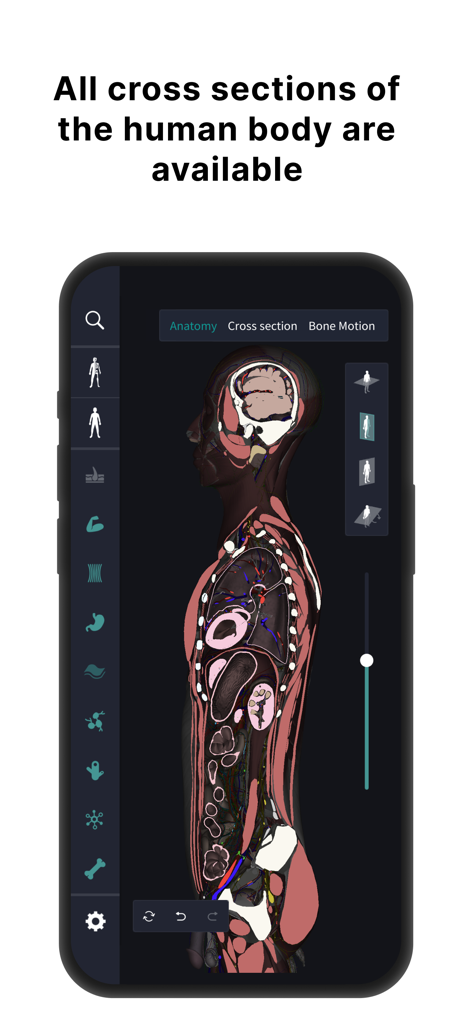
Visão de secção transversal do corpo humano no aplicativo de anatomia 3D teamLab Body Pro.
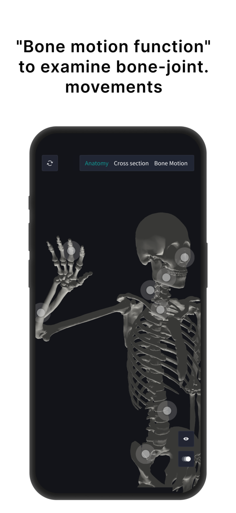
Modelo 3D do esqueleto humano no teamLab Body Pro mostrando pontos interativos de movimento articular
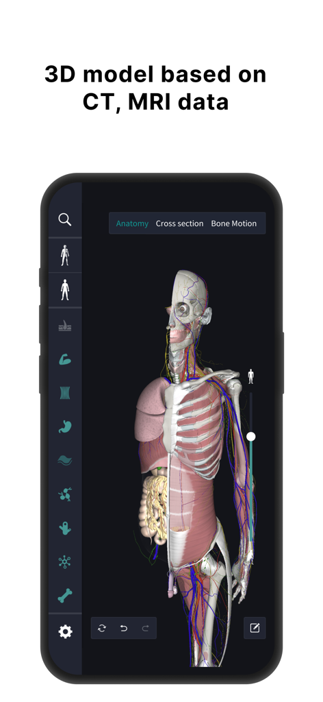
Modelo 3D de anatomia humana mostrando órgãos e músculos com base em dados de ressonância magnética
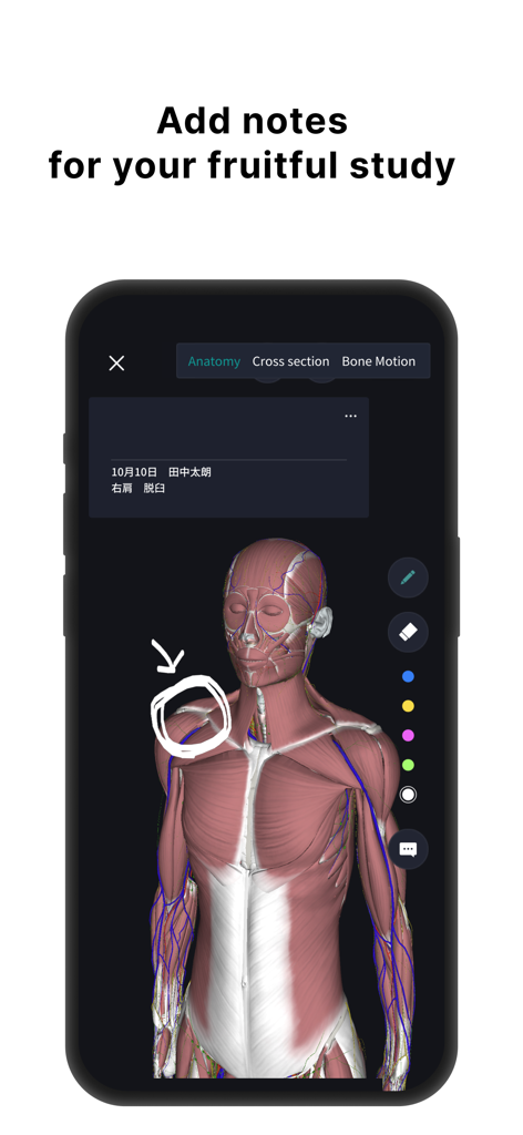
Modelo 3D de anatomia humana com camadas musculares e anotações desenhadas à mão para estudo médico.
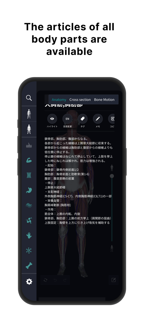
Descrições anatômicas detalhadas e interface do modelo 3D humano.
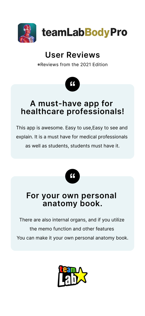
Avaliações de usuários para o aplicativo de anatomia 3D teamLab Body Pro destacando seus benefícios para profissionais de saúde e estudantes
Descrição
Download
Apps Semelhantes
Apps com recursos e experiência semelhantes

Acupuncture Points
Miridia Technology, Inc.

Anatomy by M&M
Muscle & Motion LTD

みんなde心電図
Ryoma Kamei

Histo!
John Ash

Posture by M&M
Muscle & Motion LTD

NYSORA VetRA
NYSORA Inc

Physiotutors
Physiotutors

BLOCKJOCKS
BLOCKJOCKS.COM LLC

Eastland Herb–Chinese Medicine
Eastland Press Inc.
Esta página não é uma página oficial do aplicativo ou de seu desenvolvedor, mas uma publicação editorial independente criada para fins informativos e de comentário. Salvo indicação expressa em contrário, nem o aplicativo nem seu desenvolvedor são afiliados, endossados, patrocinados, autorizados ou oficialmente conectados à MWM, Apple, Google Play, ao editor do aplicativo ou ao desenvolvedor do aplicativo, e nada nesta página implica que o aplicativo foi desenvolvido utilizando os serviços da MWM. Todas as marcas comerciais, logotipos, capturas de tela e outros conteúdos permanecem propriedade de seus respectivos proprietários.