Esta página no es una página oficial de la aplicación ni de su desarrollador, sino una publicación editorial independiente creada con fines informativos y de comentario. Salvo que se indique expresamente lo contrario, ni la aplicación ni su desarrollador están afiliados, respaldados, patrocinados, autorizados ni conectados oficialmente con MWM, Apple, Google Play, el editor de la aplicación ni su desarrollador, y nada en esta página implica que la aplicación haya sido desarrollada utilizando los servicios de MWM. Todas las marcas comerciales, logotipos, capturas de pantalla y demás contenidos son propiedad de sus respectivos propietarios.

teamLab Body Pro 3d anatomy
Mejore su práctica médica con el primer modelo 3D del mundo basado en más de 10 años de datos de resonancia magnética. Visualice el movimiento articular dinámico, realice cortes transversales personalizados y explore la estructura humana con precisión de grado quirúrgico.
Descargas
108K+Valoración
Total valoraciones
600Editor
Categoría
MedicalIdiomas
1Última versión
1.3.5Tamaño
300.9 MBFecha de lanzamiento
6 nov 2022El Estándar de Oro en Anatomía 3D
Reemplace los libros de texto estáticos con el modelo 3D más preciso del mundo, derivado de 10 años de investigación de resonancia magnética en vivo para respaldar la precisión clínica y el aprendizaje médico avanzado.
Biomecánica Articular Viva
Domine la kinesiología con las primeras animaciones 3D del mundo de movimientos articulares en vivo, que proporcionan una representación más precisa que los estudios cadavéricos tradicionales.
Corte Transversal Clínico
Genere instantáneamente planos sagitales, frontales o arbitrarios personalizados, diseñados para ayudar en el diagnóstico por ultrasonido y la planificación quirúrgica compleja.
Las siguientes capturas de pantalla y la descripción provienen directamente del listado oficial de la tienda de la aplicación y son propiedad del desarrollador.
App Store
Capturas
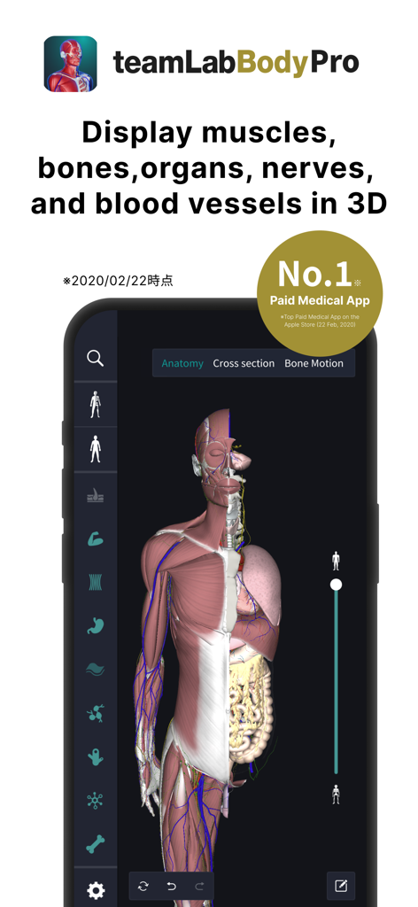
Modelo de anatomía humana 3D que muestra músculos y órganos en la aplicación teamLab Body Pro
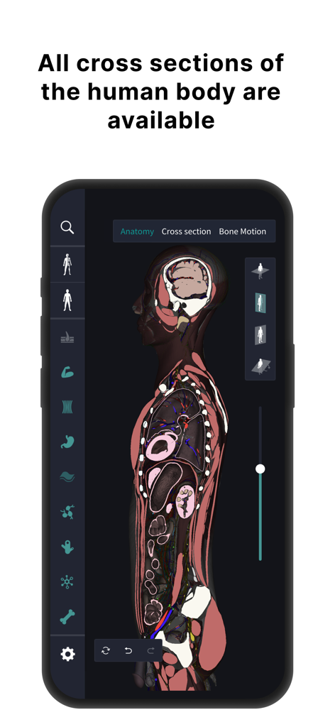
Vista de corte transversal del cuerpo humano en la aplicación de anatomía 3D teamLab Body Pro.
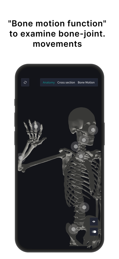
Modelo de esqueleto humano 3D en teamLab Body Pro que muestra puntos interactivos de movimiento articular
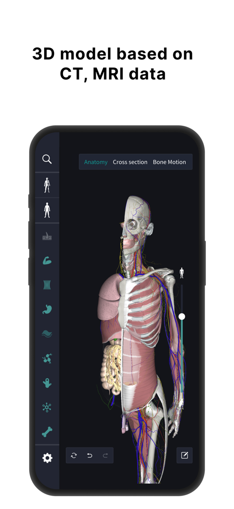
Modelo de anatomía humana 3D que muestra órganos y músculos basado en datos de resonancia magnética
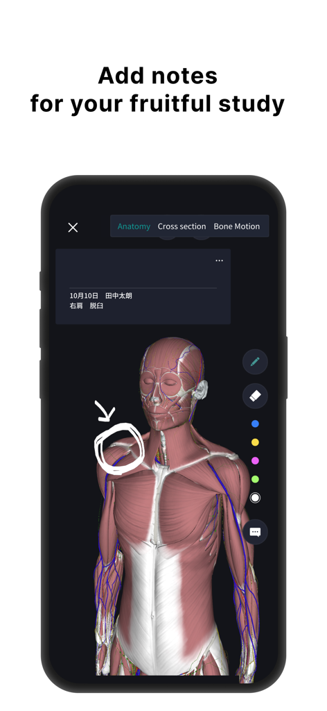
Modelo de anatomía humana 3D con capas musculares y anotaciones dibujadas a mano para el estudio médico.
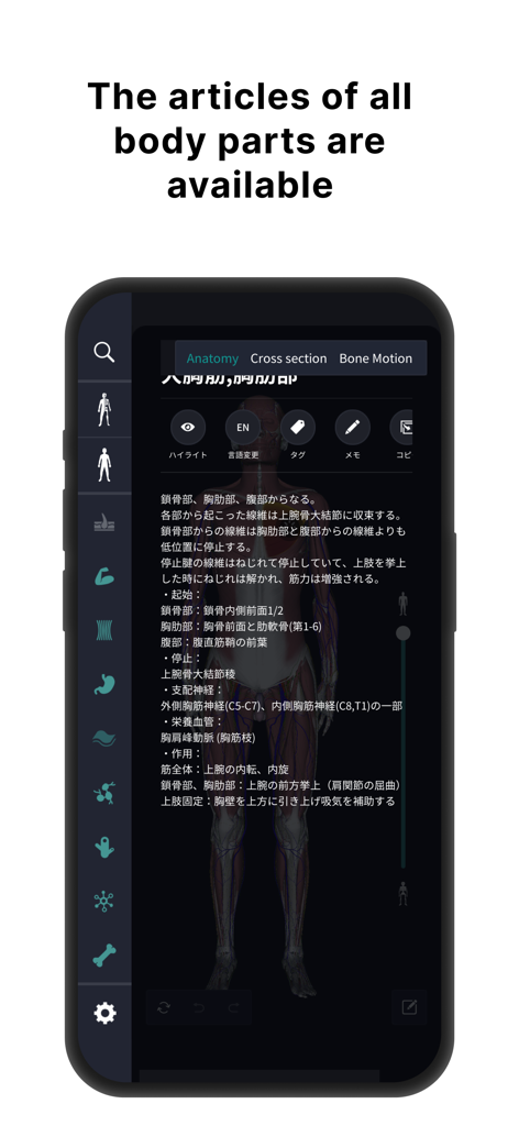
Descripciones anatómicas detalladas e interfaz de modelo humano 3D.
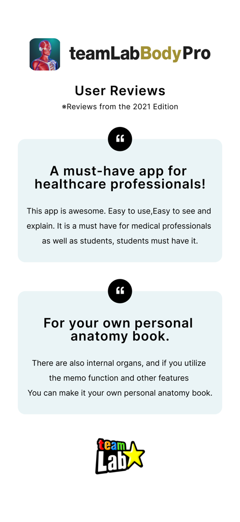
Reseñas de usuarios de la aplicación de anatomía 3D teamLab Body Pro que destacan sus beneficios para profesionales de la salud y estudiantes
Descripción
Download
Apps Similares
Apps con características y experiencia similares

Acupuncture Points
Miridia Technology, Inc.

Anatomy by M&M
Muscle & Motion LTD

みんなde心電図
Ryoma Kamei

Histo!
John Ash

Posture by M&M
Muscle & Motion LTD

NYSORA VetRA
NYSORA Inc

Physiotutors
Physiotutors

BLOCKJOCKS
BLOCKJOCKS.COM LLC

Eastland Herb–Chinese Medicine
Eastland Press Inc.
Esta página no es una página oficial de la aplicación ni de su desarrollador, sino una publicación editorial independiente creada con fines informativos y de comentario. Salvo que se indique expresamente lo contrario, ni la aplicación ni su desarrollador están afiliados, respaldados, patrocinados, autorizados ni conectados oficialmente con MWM, Apple, Google Play, el editor de la aplicación ni su desarrollador, y nada en esta página implica que la aplicación haya sido desarrollada utilizando los servicios de MWM. Todas las marcas comerciales, logotipos, capturas de pantalla y demás contenidos son propiedad de sus respectivos propietarios.