Diese Seite ist keine offizielle Seite der App oder ihres Entwicklers, sondern eine unabhängige redaktionelle Veröffentlichung, die zu Informations- und Kommentarzwecken erstellt wurde. Sofern nicht ausdrücklich anders angegeben, sind weder die App noch ihr Entwickler mit MWM, Apple, Google Play, dem App-Herausgeber oder dem Entwickler der App verbunden, von ihnen unterstützt, gesponsert, autorisiert oder anderweitig offiziell verbunden, und nichts auf dieser Seite impliziert, dass die App unter Verwendung der Dienste von MWM entwickelt wurde. Alle Marken, Logos, Screenshots und andere Inhalte bleiben Eigentum ihrer jeweiligen Inhaber.

teamLab Body Pro 3d anatomy
Verbessern Sie Ihre medizinische Praxis mit dem weltweit ersten 3D-Modell, das auf über 10 Jahren MRT-Daten basiert. Visualisieren Sie dynamische Gelenkbewegungen, führen Sie benutzerdefinierte Querschnitte durch und erkunden Sie die menschliche Struktur mit chirurgischer Präzision.
Downloads
108K+Bewertung
Bewertungen gesamt
600Herausgeber
Kategorie
MedicalSprachen
1Neueste Version
1.3.5Größe
300.9 MBErstveröffentlichung
6. Nov. 2022Der Goldstandard in der 3D-Anatomie
Ersetzen Sie statische Lehrbücher durch das weltweit genaueste 3D-Modell, das aus 10 Jahren MRT-Forschung am lebenden Körper abgeleitet wurde, um klinische Präzision und fortgeschrittene medizinische Lernprozesse zu unterstützen.
Biomechanik lebender Gelenke
Meistern Sie die Kinesiologie mit den weltweit ersten 3D-Animationen von Gelenkbewegungen am lebenden Körper, die eine genauere Darstellung als traditionelle Kadaverstudien bieten.
Klinische Querschnitte
Erstellen Sie sofort benutzerdefinierte sagittale, frontale oder beliebige Schnittebenen, die zur Unterstützung der Ultraschalldiagnostik und komplexen chirurgischen Planung entwickelt wurden.
Die folgenden Screenshots und die Beschreibung stammen direkt aus dem offiziellen Store-Eintrag der App und sind Eigentum des Entwicklers.
App Store
Screenshots
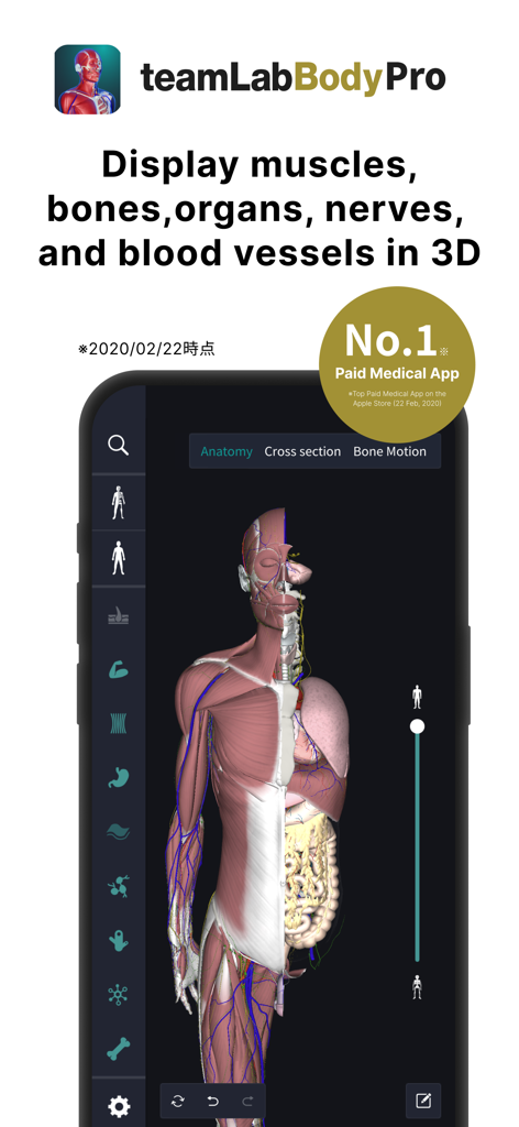
3D-Modell der menschlichen Anatomie, das Muskeln und Organe in der teamLab Body Pro App zeigt
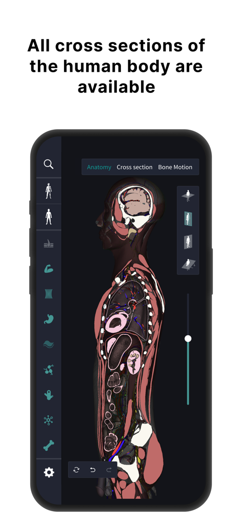
Querschnittsansicht des menschlichen Körpers in der teamLab Body Pro 3D-Anatomie-App.
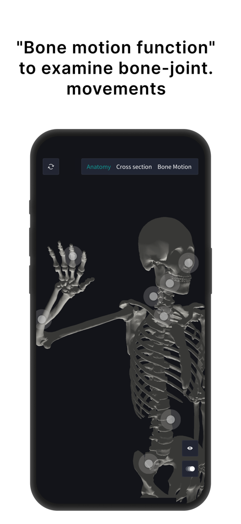
3D-Modell des menschlichen Skeletts in teamLab Body Pro, das interaktive Punkte für Gelenkbewegungen zeigt
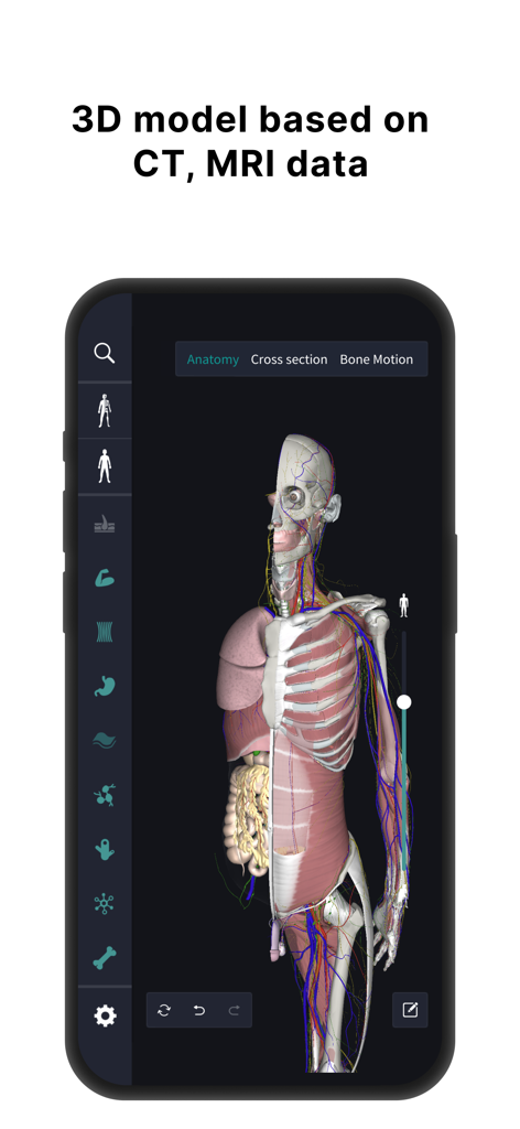
3D-Modell der menschlichen Anatomie, das Organe und Muskeln auf der Grundlage von MRT-Daten zeigt
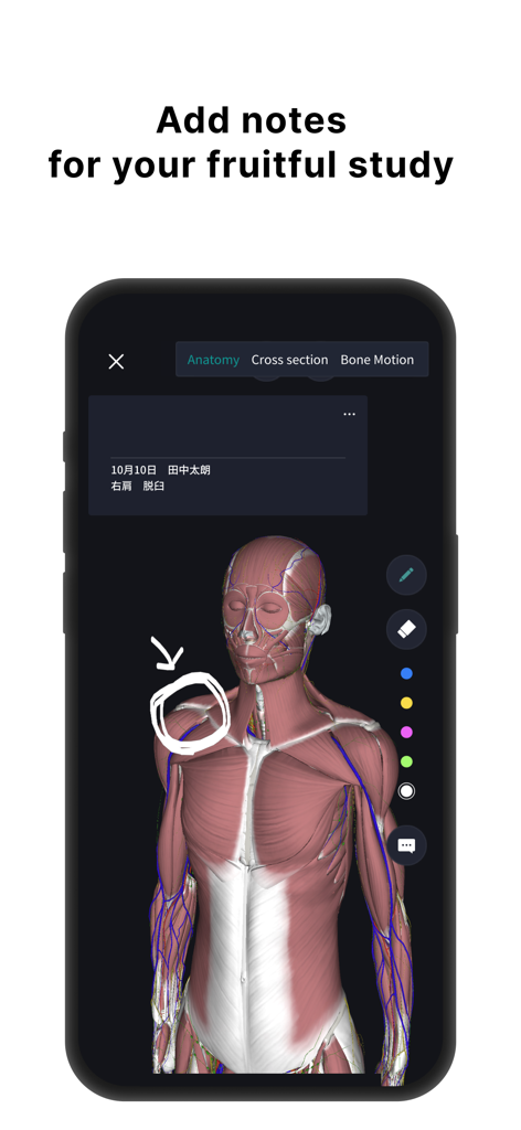
3D-Modell der menschlichen Anatomie mit Muskelschichten und handgezeichneten Anmerkungen für das Medizinstudium.
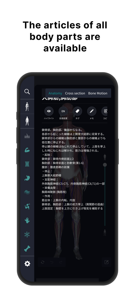
Detaillierte anatomische Beschreibungen und 3D-Mensch-Modell-Oberfläche.
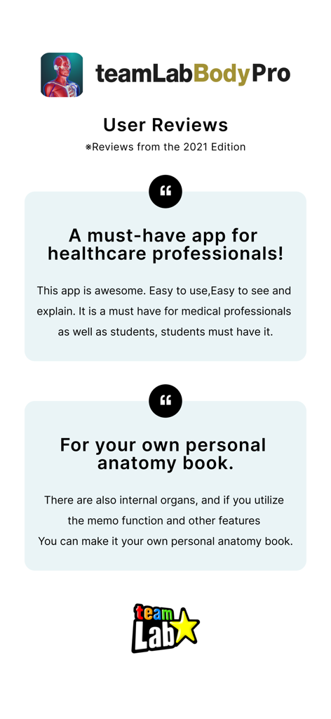
Benutzerrezensionen für die teamLab Body Pro 3D-Anatomie-App, die ihre Vorteile für medizinisches Fachpersonal und Studenten hervorheben
Beschreibung
Download
Ähnliche Apps
Apps mit ähnlichen Funktionen und Nutzererfahrung

Acupuncture Points
Miridia Technology, Inc.

Anatomy by M&M
Muscle & Motion LTD

みんなde心電図
Ryoma Kamei

Histo!
John Ash

Posture by M&M
Muscle & Motion LTD

NYSORA VetRA
NYSORA Inc

Physiotutors
Physiotutors

BLOCKJOCKS
BLOCKJOCKS.COM LLC

Eastland Herb–Chinese Medicine
Eastland Press Inc.
Diese Seite ist keine offizielle Seite der App oder ihres Entwicklers, sondern eine unabhängige redaktionelle Veröffentlichung, die zu Informations- und Kommentarzwecken erstellt wurde. Sofern nicht ausdrücklich anders angegeben, sind weder die App noch ihr Entwickler mit MWM, Apple, Google Play, dem App-Herausgeber oder dem Entwickler der App verbunden, von ihnen unterstützt, gesponsert, autorisiert oder anderweitig offiziell verbunden, und nichts auf dieser Seite impliziert, dass die App unter Verwendung der Dienste von MWM entwickelt wurde. Alle Marken, Logos, Screenshots und andere Inhalte bleiben Eigentum ihrer jeweiligen Inhaber.