This page is not an official page of the app or its developer, but an independent editorial publication created for informational and commentary purposes. Unless expressly stated otherwise, neither the app nor its developer is affiliated with, endorsed by, sponsored by, authorized by, or otherwise officially connected with MWM, Apple, Google Play, the app publisher, or the app's developer, and nothing on this page implies that the app was developed using MWM's services. Any trademarks, logos, screenshots, and other content remain the property of their respective owners.

teamLab Body Pro 3d anatomy
Elevate your medical practice with the world’s first 3D model based on 10+ years of MRI data. Visualize dynamic joint movement, perform custom cross-sections, and explore human structure with surgical-grade precision.
Downloads
108K+User Rating
Total Ratings
600Publisher
Category
MedicalLocales
1Latest Version
1.3.5Size
300.9 MBFirst Released
Nov 6, 2022The Gold Standard in 3D Anatomy
Replace static textbooks with the world's most accurate 3D model, derived from 10 years of live MRI research to support clinical precision and advanced medical learning.
Living Joint Biomechanics
Master kinesiology with the world's first 3D animations of live joint motion, providing a more accurate representation than traditional cadaveric studies.
Clinical Cross-Sectioning
Instantly generate custom sagittal, frontal, or arbitrary planes, designed to assist in ultrasound diagnosis and complex surgical planning.
The following screenshots and description are sourced directly from the app's official store listing and are the property of the app developer.
App Store
Screenshots
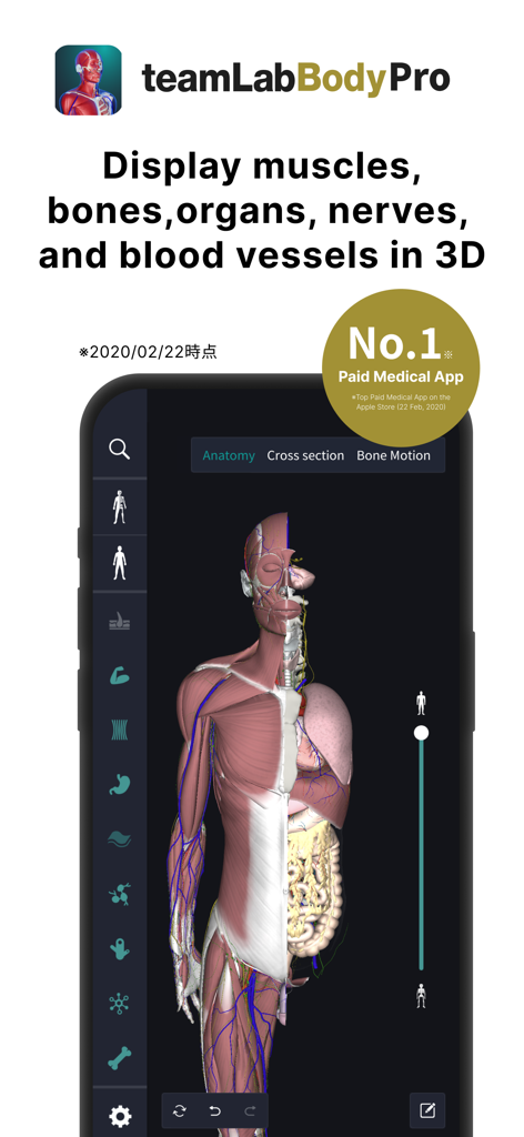
3D human anatomy model showing muscles and organs in the teamLab Body Pro app
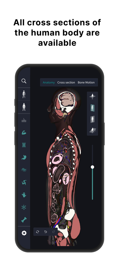
Human body cross section view in the teamLab Body Pro 3D anatomy app.
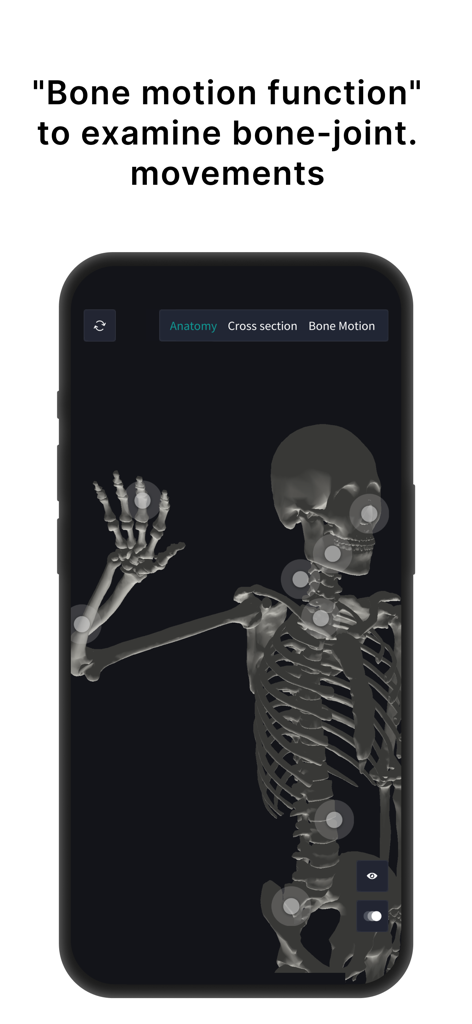
3D human skeleton model in teamLab Body Pro showing joint movement interactive points
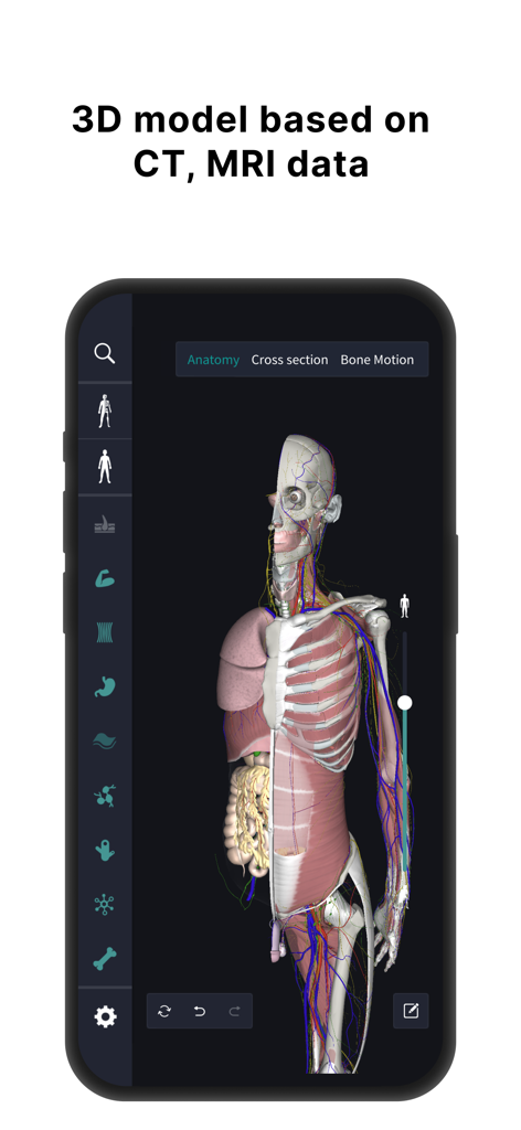
3D human anatomy model showing organs and muscles based on MRI data
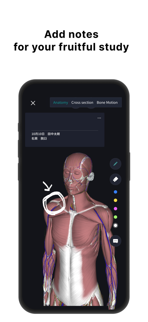
3D human anatomy model with muscle layers and hand-drawn annotations for medical study.
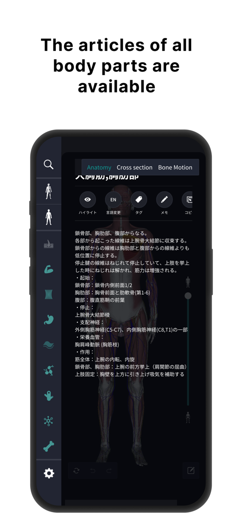
Detailed anatomical descriptions and 3D human model interface.
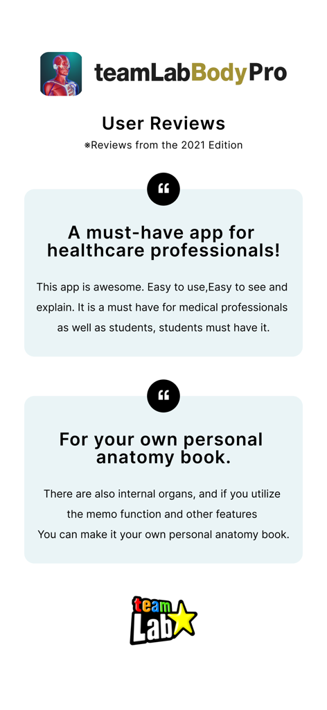
User reviews for teamLab Body Pro 3D anatomy app highlighting its benefits for healthcare professionals and students
Description
Download
More Like This
Apps with similar features and user experience

Acupuncture Points
Miridia Technology, Inc.

Anatomy by M&M
Muscle & Motion LTD

みんなde心電図
Ryoma Kamei

Histo!
John Ash

Posture by M&M
Muscle & Motion LTD

NYSORA VetRA
NYSORA Inc

Physiotutors
Physiotutors

BLOCKJOCKS
BLOCKJOCKS.COM LLC

Eastland Herb–Chinese Medicine
Eastland Press Inc.
This page is not an official page of the app or its developer, but an independent editorial publication created for informational and commentary purposes. Unless expressly stated otherwise, neither the app nor its developer is affiliated with, endorsed by, sponsored by, authorized by, or otherwise officially connected with MWM, Apple, Google Play, the app publisher, or the app's developer, and nothing on this page implies that the app was developed using MWM's services. Any trademarks, logos, screenshots, and other content remain the property of their respective owners.